Nematodiasis in laying ducks (Anas platyrhynchos) at different age groups
DOI:
https://doi.org/10.46549/jipvet.v15i4.586Keywords:
Nematode infection, Age, Occurrence , Gastrointestinal parasites, Laying ducksAbstract
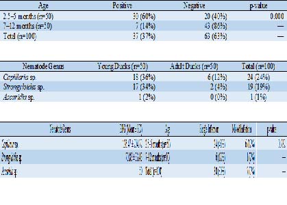
Nematodiasis remains a critical parasitic disease affecting the gastrointestinal health and productivity of laying ducks. This study aimed to investigate the occurrence of nematode infections at different ages in laying ducks (Anas platyrhynchos). A total of 100 intensively managed local ducks were used, comprising 50 young ducks (2.5–5 months) and 50 adults (7 months–1 year). Fecal samples were analyzed using the modified Whitlock flotation technique. Descriptive analysis and Chi-square statistical testing were performed using SPSS software. The overall occurrence of nematodiasis was 37% (37/100), with Capillaria sp. being the most frequently detected species (24%), followed by Strongyloides sp. (19%) and Ascaridia sp. (1%). Single-species infections predominated (81.1%), while mixed-species infections accounted for 18.9% of positive cases. Eggs per gram (EPG) values varied among age groups but were consistently within the mild infection range. Statistical analysis revealed a significant association between age group and infection occurrence (p<0,05), indicating that younger ducks are at higher risk of nematode infection. These findings highlight the importance of age-specific parasite control strategies to enhance flock health and productivity in laying duck operations.
Downloads
References
Adejinmi JO, Oke M. 2011. Gastro-intestinal parasites of domestic ducks (Anas platyrhynchos) in Ibadan Southwestern Nigeria. Asian Journal of Poultry Science. 5(1); 46-50. https://doi.org/10.3923/ajpsaj.2011.46.50
Anggrahini S, Widiyono I, Baihaqi ZA, Sofyan A, Mulianda R, Wulandari W, Ekawasti F, Fauziah I, Sadarman S, Sigit M, Herdian H, Ahmad RZ, Rokana E. 2025. Occurrence of gastrointestinal parasites in local ducks at varying altitudes in Yogyakarta, Indonesia. Veterinary World. 18(3); 616-623. https://doi.org/10.14202/vetworld.2025.616-623
Arlina F, Husmaini S, Rhoudha R, Sardi WR, Rafian T. 2021. Keragaman fenotipe kualitatif dan kuantitatif itik Sikumbang Jonti sebagai plasma nutfah di Sumatera Barat. Jurnal Ilmu Peternakan dan Veteriner Tropis. 11(3); 291-299. https://doi.org/10.46549/jipvet.v11i3.173
Bajoi MSJ, Bhutto B, Soomro F, Solangi IA, Marri NU, Kakar ZK, Bangulzai M, Baloch AL, Mangrio RA, Khosa TUD, Mengal MA, Marri NM, Kabir A. 2024. Prevalence and characteristics of gastrointestinal parasites in backyard chickens of Khuzdar, Baluchistan. Journal of Survey in Fisheries Sciences. 11(4); 63-69. https://doi.org/10.53555/sfs.v11i4.2742
Ballweber LR. 2001. Veterinary Parasitology. USA: Butterworth-Heinemann.
Carvalho EL, Santana RLS, Sousa DF, Cabral GS, Pinheiro RHS, Pereira WLA, Giese EG. 2021. Lesions caused by anisakid and capillariid in Cairina moschata raised on Marajó island, state of Pará, Brazil. Arquivo Brasileiro de Medicina Veterinária e Zootecnia. 73(4); 885-892. https://doi.org/10.1590/1678-4162-12334
Dalal D, Saha S, Dalal DD, Roy A, Talapatra SN, Ghosh P. 2024. Comparative study on the outbreak of helminth parasites in domestic duck and chicken. International Journal of Fauna and Biological Studies. 11(2); 23-26. https://doi.org/10.22271/23940522.2024.v11.i2a.1015
El-Ghany WA. 2022. An updated insight into the gastrointestinal helminthoses of poultry: a review. Annals of Parasitology. 68(4); 645-656. https://doi.org/10.17420/ap6804.471
Hasanien NE, Dyab AK, Arafa MI, Nasr AAE, Mohamed SAA. 2025. Prevalence of intestinal parasites of domestic duck in Assiut, Egypt: with special reference for coccidian infection. Assiut Veterinary Medical Journal. 71(184); 364-375. https://doi.org/10.21608/avmj.2024.342749.1514
Henrik, Marhayani, Syadik F. 2021. Karakteristik morfometrik itik dan produksi telur itik di sentra peternakan itik Kabupaten Tolitoli. Jurnal Ilmu Peternakan dan Veteriner Tropis. 11(3); 204-210. https://doi.org/10.46549/jipvet.v11i3.189
Makouloutou-Nzassi P, Longo-Pendy NM, Nguema LKA, Lendzele SS, Bangueboussa F, Bouchedi B, Maganga GD, Boundenga L. 2024. Prevalence of gastrointestinal parasites in chickens (Gallus gallus domesticus) and associated risk factors in M'passa department, Southeast Gabon. Open Veterinary Journal. 14(12); 3232-3240. https://doi.org/10.5455/OVJ.2024.v14.i12.8
Martawijaya EI, Martanto E, Tinaprilla N. 2004. Panduan Beternak Itik Petelur Secara Intensif. Jakarta: AgroMedia Pustaka.
Maulana H. 2023. Beternak Itik Petelur. Jakarta: AgroMedia.
Muhairwa AP, Msoffe PL, Ramadhani S, Mollel EL, Mtambo MA, Kassuku AA. 2007. Prevalence of gastro-intestinal helminths in free-range ducks in Morogoro Municipality, Tanzania. Livestock Research for Rural Development. 19(4); 1-6. https://www.lrrd.org/lrrd19/4/muha19048.html
Nurtjahyani SD, Agustin DS. 2014. Prevalensi infeksi telur cacing nematoda pada feses sapi potong (Bos sp) dengan metode Whitlock. Seminar Nasional XI Pendidikan Biologi FKIP UNS; 539-543.
Ola-Fadunsin SD, Uwabujo PI, Sanda IM, Ganiyu IA, Hussain K, Rabiu M, Elelu N, Alayande MO. 2019. Gastrointestinal helminths of intensively managed poultry in Kwara Central, Kwara State, Nigeria: its diversity, prevalence, intensity, and risk factors. Veterinary World. 12(3); 389-396. https://doi.org/10.14202/vetworld.2019.389-396
Onyeabor AI, Onunkwo DN. 2023. Preliminary survey on the intestinal protozoan and helminthes parasites of domestic ducks reared in Umuahia Area of Abia State. The International Journal of Agriculture, Management and Technology. 7(1); 637-640. www.ijamt.com.ng
Otranto D, Wall R. 2024. Veterinary Parasitology. 5th ed. UK: Wiley Blackwell. https://doi.org/10.1002/9781394176373
Pepper CM, Dunlop MW. 2021. Review of litter turning during a grow-out as a litter management practice to achieve dry and friable litter in poultry production. Poultry Science. 100(6). https://doi.org/10.1016/j.psj.2021.101071
Permatasari DA, Rochiman K, Restiadi TI, S MS, Suprihati E, Effendi MH. 2020. Prevalence and worm infection degree gastrointestinal on duck (Anas javanica) in two different geographical territory. Journal of Parasite Science. 4(1); 21-24. https://doi.org/10.20473/jops.v4i1.20271
Salsabila N, Bekti NS, Widiyono I. 2023. Nematode and coccidia infections in singing birds kept in bird stalls. IOP Conference Series: Earth and Environmental Science. 1174(1); 012014. https://doi.org/10.1088/1755-1315/1174/1/012014
Shemshadi B, Ranjbar-bahadori S, Delfan-abazari M. 2017. Prevalence and intensity of parasitic infection in domestic ducks (Anas platyrhynchas) in Gilan Province, Northern Iran. Comparative Clinical Pathology. 26(1); 165-167. https://doi.org/10.1007/s00580-016-2361-7
Shrestha D, Subedi JR, Chhetri B. 2020. Gastrointestinal parasites of domesticated duck (Anas platyrhynchos Linnaeus, 1758) in Chandragiri municipality, Kathmandu, Nepal. Ife Journal of Science. 22(2); 015-023. https://doi.org/10.4314/ijs.v22i2.2
Temesgen AB, Wassie ZG, Abebe S. 2024. Prevalence of GIT nematodes and associated risk factors of exotic chickens in selected farm of poultry in and around Ambo, Ethiopia. BioRxiv; 78-91. https://doi.org/10.1101/2024.07.02.601802
Thienpont D, Rochette D, Vanparijs OFJ. 2003. Diagnosing Helminthiasis by Coprological Examination. 3rd ed. Belgium: Janssen Animal Health. https://www.researchgate.net/publication/283924775
Thrusfield M. 2007. Veterinary Epidemiology. 3rd ed. Oxford: Blackwell Publishing.
Tiersch KM, Daş G, Samson-Himmelstjerna GV, Gauly M. 2013. The role of culture media on embryonation and subsequent infectivity of Capillaria obsignata eggs. Parasitology Research. 112(1); 357-364. https://doi.org/10.1007/s00436-012-3143-z
Wakhid A. 2023. Petunjuk Praktis Beternak Itik Petelur. Jakarta: AgroMedia.
Waruiru RM, Mavuti SK, Mbuthia PG, Njagi LW. 2018. Prevalence and intensity of gastrointestinal helminth infestations of free range domestic ducks in Kenya. Livestock Research for Rural Development. 30(4). https://www.lrrd.org/lrrd30/4/rmwa30066.html
Widiyono I, Rusmihayati, Purnamaningsih H, Sahara A. 2023. Zoonotic gastrointestinal nematodes in pet cats in Yogyakarta (Indonesia) and their susceptibility to anthelmintics. Biodiversitas; 3332-3337. https://doi.org/10.13057/biodiv/d240628
Win SY, Htun LL, Hmoon MM, Chel HM, Thaw YN, Soe NC, Oo HL, Bawm S. 2020. Occurrence of gastrointestinal parasites in free ranging village chickens from four townships of Myanmar. Veterinary Sciences: Research and Reviews. 6(1). https://doi.org/10.17582/journal.vsrr/2020/6.1.1.6
Yuriwati FN, Mardiati SM, Tana S. 2016. Perbandingan struktur histologi magnum pada itik Magelang, itik Tegal dan itik Pengging. Buletin Anatomi dan Fisiologi. 24(1); 76-85.
Zaharah I, Yanti AH, Setyawati TR. 2016. Kepadatan nematoda gastrointestinal itik Manila (Cairina moschata) yang dipasarkan di pasar Flamboyan Kota Pontianak. Protobiont. 5(3); 41-46.
Downloads
Published
How to Cite
Issue
Section
License
Copyright (c) 2025 Intania N. Khasanah , Irkham Widiyono

This work is licensed under a Creative Commons Attribution-NonCommercial-ShareAlike 4.0 International License.
License and Copyright Agreement
In submitting the manuscript to the journal, the authors certify that:
- They are authorized by their co-authors to enter into these arrangements.
- The work described has not been formally published before, except in the form of an abstract or as part of a published lecture, review, thesis, or overlay journal. Please also carefully read Jurnal Ilmu Peternakan dan Veteriner Tropis (Journal of Tropical Animal and Veterinary Science) Posting Your Article Policy at https://journal.fapetunipa.ac.id/index.php/JIPVET/publicationethics
- That it is not under consideration for publication elsewhere,
- That its publication has been approved by all the author(s) and by the responsible authorities “tacitly or explicitly“ of the institutes where the work has been carried out.
- They secure the right to reproduce any material that has already been published or copyrighted elsewhere.
- They agree to the following license and copyright agreement.
Copyright
Authors who publish with Jurnal Ilmu Peternakan dan Veteriner Tropis (Journal of Tropical Animal and Veterinary Science) agree to the following terms:
- Authors retain copyright and grant the journal right of first publication with the work simultaneously licensed under a Creative Commons Attribution License (CC BY-NC-SA 4.0) that allows others to share the work with an acknowledgment of the work's authorship and initial publication in this journal.
- Authors are able to enter into separate, additional contractual arrangements for the non-exclusive distribution of the journal's published version of the work (e.g., post it to an institutional repository or publish it in a book), with an acknowledgment of its initial publication in this journal.
- Authors are permitted and encouraged to post their work online (e.g., in institutional repositories or on their website) prior to and during the submission process, as it can lead to productive exchanges, as well as earlier and greater citation of published work.

This work is licensed under a Creative Commons Attribution-NonCommercial-ShareAlike 4.0 International License.


.png)
